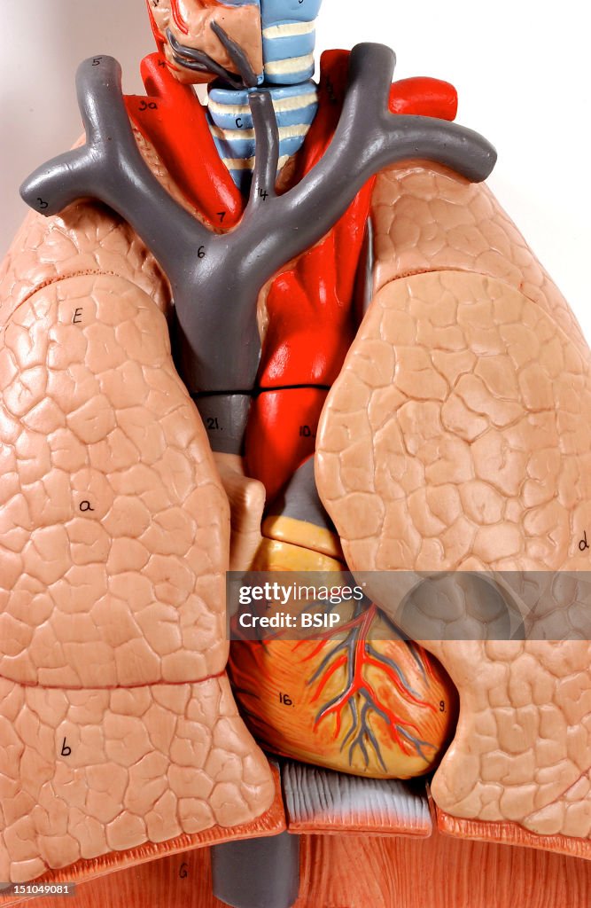
Lung, Anatomy
Anatomic Model Of The Chest Organs Trachea, Lung, Heart Of An Adult Human Body Anterior View. At The Throat Level, The Cartilaginous Rings Of The Trachea Are Depicted In Light Blue. The Right Side Of The Neck Shows The Thyroid Gland In Nude Covering The Trachea. The Lungs, Wrapped By The Pleura, Are Constituted By Multiple Compartments, The Lobules. The Right And Left Lungs Are Each Composed Of An Upper And A Lower Lobes B And D, Separated By An Oblique Fissure. The Right Lung Has An Additional Lobe, The Middle Lobe A, That Is Inferiorly Demarcated By The Horizontal Fissure. The Left Lobe Has An Indentation, The Cardiac Notch, In Which Lies The Heart. The Heart Contains Four Cavities: Two Atriums In Its Upper Part And Two Ventricles In Light Orange, Wandered By The Coronary Circulation In Its Lower Part. The Inferior Thyroid Vein In Black Emerges From The Thyroid Gland And Lead To The Internal Jugular Vein. The Right And Left Internal Jugular Veins Begin At The Base Of The Skull, Go Down On Both Sides Of The Throat To Reach The Clavicles, Where They Join Up With The Subclavian Veins Coming From The Upper Limbs To Form The Brachiocephalic Trunk. The Latter Flows Into The Superior Vena Cava In Black, Which Brings To The Heart The Deoxygenated Blood Coming From The Upper Part Of The Body. The Blood Is Next Oxygenated By The Lungs And Propeled By The Heart Towards The Whole Body Through The Aorta In Red. The Ascending Aorta Divides First Into The Brachiocephalic Trunk, Then Into The Common Carotid Arteries, Vascularizing Neck And Head Structures, And Lastly Into The Subclavian Arteries, Irrigating The Upper Limbs. The Diaphragm Separates The Thorax From The Abdominal Cavity. (Photo By BSIP/UIG Via Getty Images)

COMPRAR LICENCIA
¿Cómo puedo usar esta imagen?
385,00 €
EUR
Getty ImagesLung, Anatomy, Fotografía de noticias Lung, Anatomy Consigue fotografías de noticias de alta resolución y gran calidad en Getty ImagesProduct #:151049081
 Lung, Anatomy Consigue fotografías de noticias de alta resolución y gran calidad en Getty ImagesProduct #:151049081
475€175€
Getty Images
In stockTen en cuenta lo siguiente: las imágenes que representan eventos históricos pueden contener temas, o tener descripciones, que no reflejan la comprensión actual. Se proporcionan en un contexto histórico. Más información.
DETALLES
Restricciones:
Póngase en contacto con su oficina local para conocer todos los usos con fines comerciales o promocionales.
Crédito:
Editorial n.º:
151049081
Colección:
Universal Images Group
Fecha de creación:
23 de junio de 2005
Fecha de subida:
Tipo de licencia:
Inf. de autorización:
No se cuenta con autorizaciones. Más información
Fuente:
Universal Images Group Editorial
Nombre del objeto:
941_04_1147805
Tamaño máx. archivo:
2365 x 3630 px (20,02 x 30,73 cm) - 300 dpi - 2 MB
- Pulmón,
- Anatomía,
- Adulto,
- Aorta,
- Aorta ascendente,
- Arteria,
- Arteria carótida,
- Arteria subclavicular,
- Cardiólogo,
- Equipo respiratorio,
- Flujo sanguíneo,
- Fondo con color,
- Incisura cardíaca,
- Interés humano,
- Laringe,
- Latido cardíaco,
- Lóbulo - Descripción física,
- Lóbulo medio,
- Neumología,
- Pericardio,
- Personas,
- Pleura,
- Plástico,
- Sangre,
- Sistema cardiovascular,
- Sistema respiratorio,
- Tráquea - Vía respiratoria,
- Vaso sanguíneo,
- Vena Cava - Vena humana,
- Vena cardíaca,
- Vena subclavicular,
- Vena yugular,
- Vena yugular interna,
- Vertical,
- Vista de frente,
- Órgano interno humano,