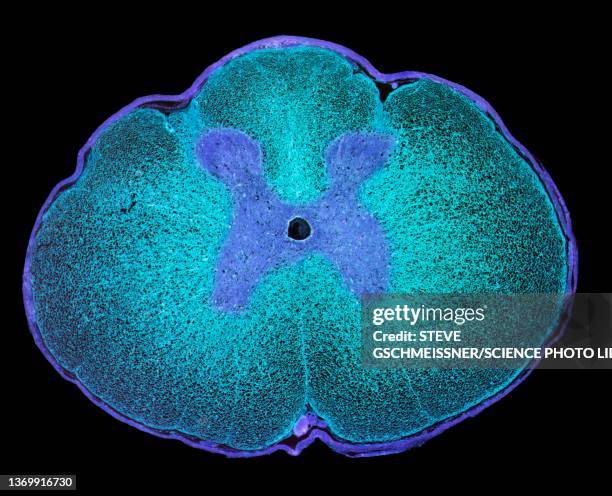
hippocampal sulcus remnant cysts seen on t2 axial mri (magnetic resonance imaging) - cerebral cortex fotografías e imágenes de stock
cortex of ranunculus root. parenchyma tissue with starch grains intercellular space. 100x - cerebral cortex fotografías e imágenes de stock
Illustration of the location of the black substance, blue). This area contains dopamine neurons that are produced in smaller quantities in the case...
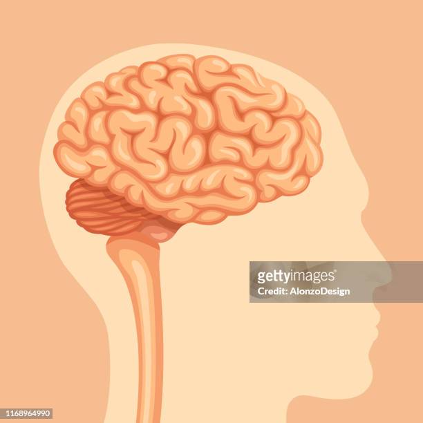
ilustraciones, imágenes clip art, dibujos animados e iconos de stock de cerebellum, illustration - cerebral cortex
Cerebral atrophy, anterior temporal and parietal frontal Ponto cerebellar, saggital plane MRI cranial scan.
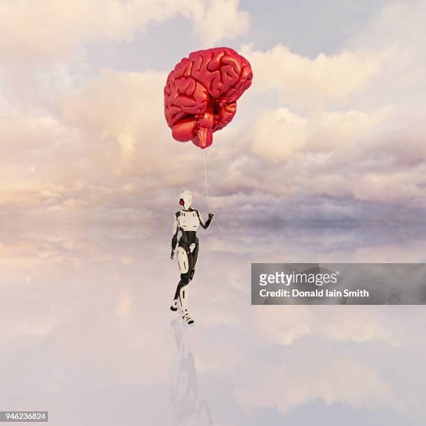
female android holds large floating human brain like balloon on a string - cerebral cortex fotografías e imágenes de stock
ilustraciones, imágenes clip art, dibujos animados e iconos de stock de hippocampus neuron, illustration - cerebral cortex
human versus machine: robot supplicates before giant floating human brain - cerebral cortex fotografías e imágenes de stock
ilustraciones, imágenes clip art, dibujos animados e iconos de stock de microaneurysms, illustration - cerebral cortex
ilustraciones, imágenes clip art, dibujos animados e iconos de stock de pyramidal neurons in the cerebral cortex, illustration - cerebral cortex
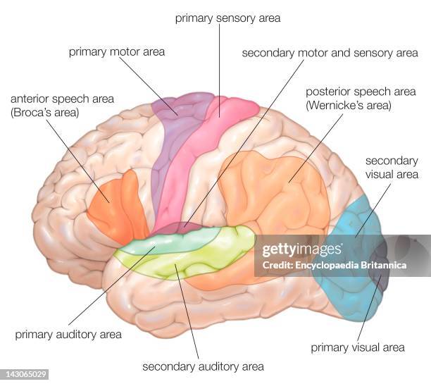
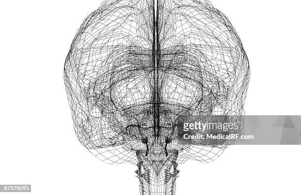
ilustraciones, imágenes clip art, dibujos animados e iconos de stock de anatomía del cerebro humano. vista frontal - cerebral cortex
Cerebral atrophy, anterior temporal and parietal frontal Ponto cerebellar, radial cross-section MRI cranial scan.
Cerebral atrophy, anterior temporal and parietal frontal Ponto cerebellar, radial cross-section MRI cranial scan.
Cerebral atrophy, anterior temporal and parietal frontal Ponto cerebellar, frontal cross-section MRI cranial scan.
Cerebral atrophy, anterior temporal and parietal frontal Ponto cerebellar, frontal cross-section MRI cranial scan.
ilustraciones, imágenes clip art, dibujos animados e iconos de stock de human brain anatomy, 3d illustration - cerebral cortex
ilustraciones, imágenes clip art, dibujos animados e iconos de stock de protoplasmic and fibrous astrocytes, illustration - cerebral cortex
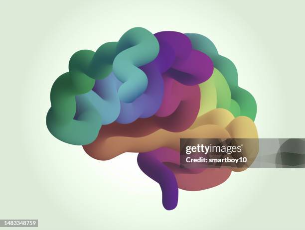
ilustraciones, imágenes clip art, dibujos animados e iconos de stock de cerebro humano estilizado - cerebral cortex
Photo Essay From Hospital. Grenoble University Hospital Michallon Hospital Department Of Neurology Unit 'Movement Disorder'. Mri Of A 30 Year Old...
Neuron, Pyramidal Neurons Of Cat Cerebral Cortex, Each Neuron Is Composed Of A Cell Body And Different Prolongations, An Axon And Several Dendrites,...
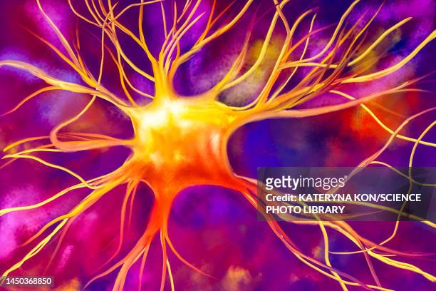
ilustraciones, imágenes clip art, dibujos animados e iconos de stock de nerve cell, illustration - cerebral cortex
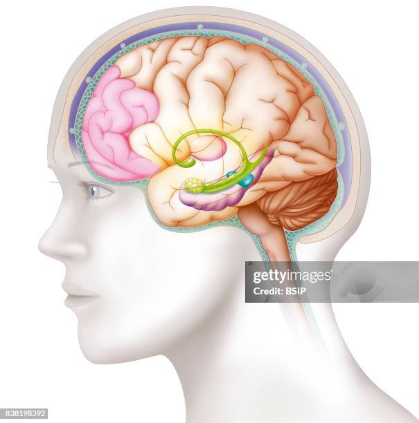
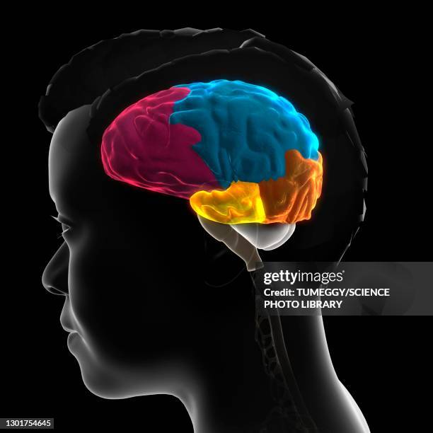
Illustration of the areas in the brain involved in the process of depression. Lessening in volume and activity of the prefrontal cortex and the...
Cerebral atrophy, anterior temporal and parietal frontal Ponto cerebellar, frontal cross-section MRI cranial scan.
Cerebral atrophy, anterior temporal and parietal frontal Ponto cerebellar, frontal cross-section MRI cranial scan.
Cerebral atrophy, anterior temporal and parietal frontal Ponto cerebellar, radial cross-section MRI cranial scan.
ilustraciones, imágenes clip art, dibujos animados e iconos de stock de anatomía del cerebro humano, publicado en 1876 - cerebral cortex
Sagittal Section. Cf. Image 0212106 For The Numbers1. Brain. 2. Corpus Callosum Splenium. 3. Septum Lucidum. 4. Thalamus. 5. Mamillary Body. 6....
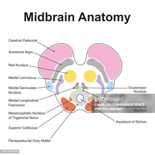
ilustraciones, imágenes clip art, dibujos animados e iconos de stock de midbrain anatomy, illustration - cerebral cortex
rathke cleft cysts, also known as pars intermedia cysts seen on sagittal mri t1 postgadolinium image - cerebral cortex fotografías e imágenes de stock
ilustraciones, imágenes clip art, dibujos animados e iconos de stock de pyramidal neurons and myelin sheaths, illustration - cerebral cortex
ilustraciones, imágenes clip art, dibujos animados e iconos de stock de dorsal striatum, illustration - cerebral cortex
ilustraciones, imágenes clip art, dibujos animados e iconos de stock de human brain, illustration - cerebral cortex
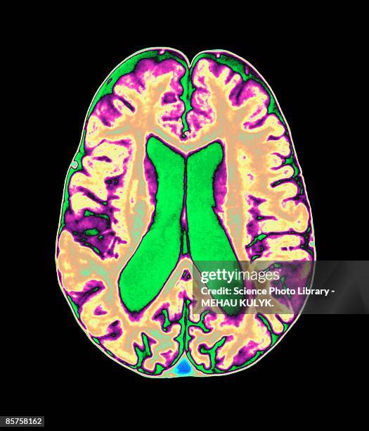
la tomografía computarizada del cerebro y la resonancia magnética del cerebro le permiten obtener una imagen tridimensional de varias partes del cerebro humano, incluida la corteza cerebral, sus partes internas y externas. - cerebral cortex fotografías e imágenes de stock
ilustraciones, imágenes clip art, dibujos animados e iconos de stock de elemento de diseño de línea cerebral humana - cerebral cortex
ilustraciones, imágenes clip art, dibujos animados e iconos de stock de human nervous system, illustration - cerebral cortex
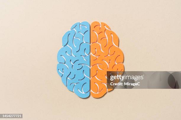
ilustraciones, imágenes clip art, dibujos animados e iconos de stock de human brain cerebral hemispheres, illustration - cerebral cortex
ilustraciones, imágenes clip art, dibujos animados e iconos de stock de anatomía del cerebro humano - cerebral cortex
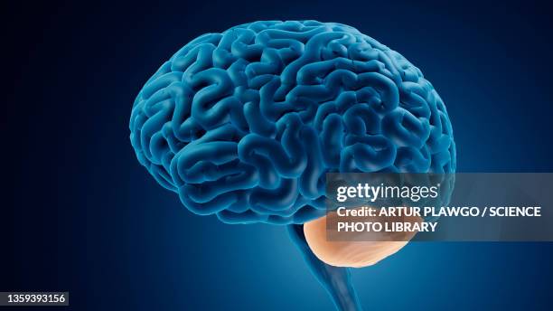
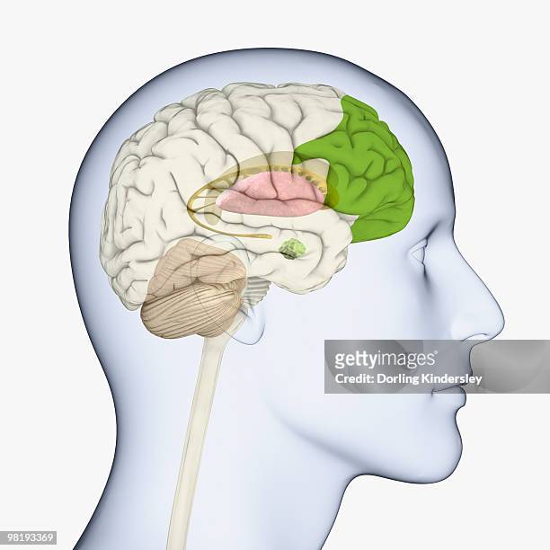
ilustraciones, imágenes clip art, dibujos animados e iconos de stock de digital illustration of head in profile showing brain of mid adult man with fully developed prefrontal cortex - cerebral cortex