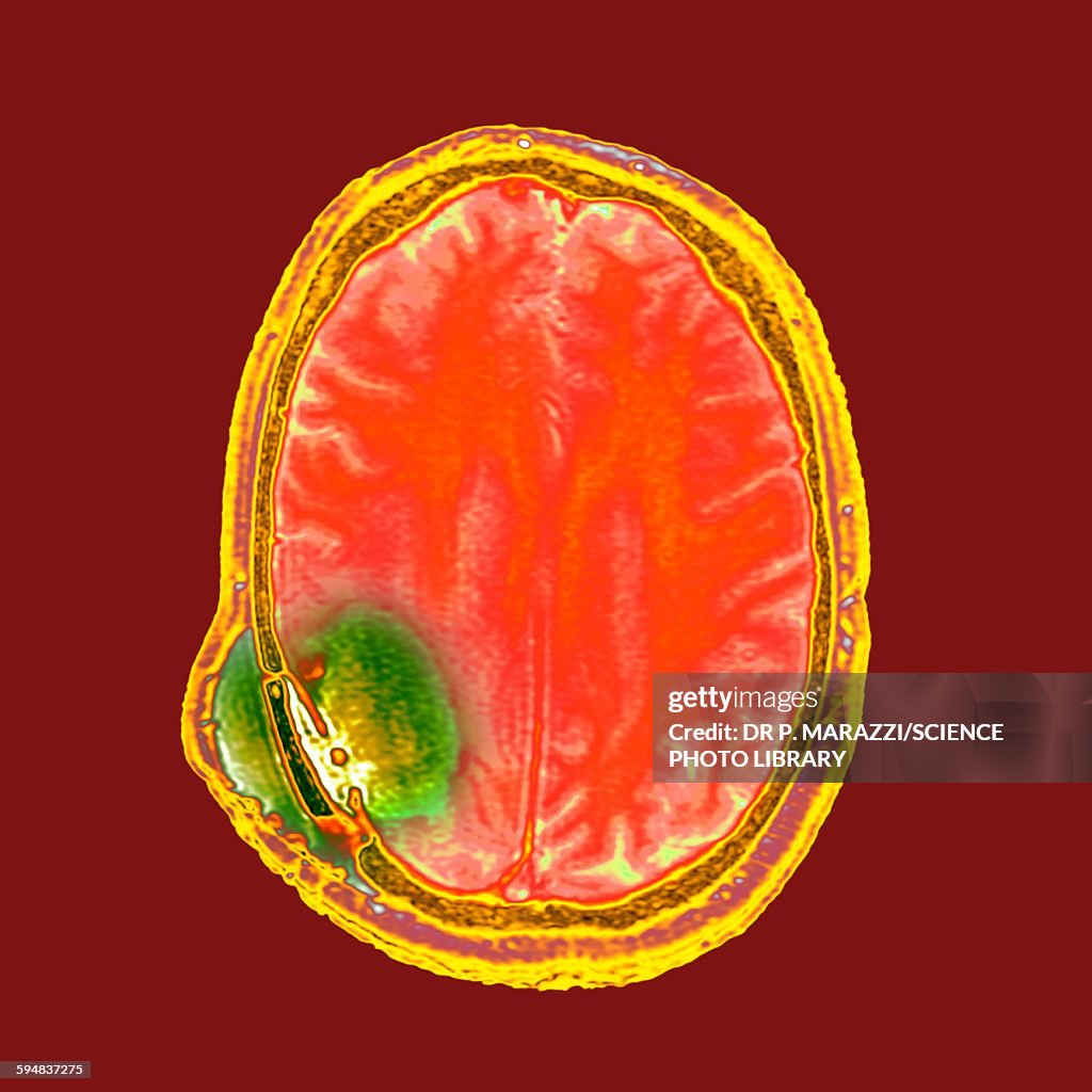
Brain cancer after surgery, MRI scan - Fotografía de stock
Brain cancer after surgery. Coloured magnetic resonance imaging (MRI) scan of a section through the head of a 59-year-old male patient with glioblastoma multiforme, showing some improvement in the disease affecting the white matter after surgery. Glioblastoma multiforme is a malignant (cancerous) tumour that arises from astrocytes, one of the support cells of the brain. It is an aggressive cancer with a poor prognosis. Here, the cancer in the right temporo-parietal cavity (lower right) has largely improved. However, a small lesion is still evident and a new area of potential disease (green) is now evident in the cortex of the occipital lobe.

COMPRAR LICENCIA
Todas las licencias libres de derechos incluyen derechos de uso mundiales, protección completa y precios sencillos con descuentos por volumen.
475,00 €
EUR
DETALLES
Creative n.º:
594837275
Tipo de licencia:
Colección:
Science Photo Library
Tamaño máx. archivo:
4500 x 4500 px (38,10 x 38,10 cm) - 300 dpi - 2 MB
Fecha de subida:
Inf. de autorización:
No se precisa autorización
Categorías:
- Imagen de resonancia magnética,
- Cerebro,
- Cáncer - Tumor,
- Cáncer de cerebro,
- Tumor,
- Escáner IRM,
- 2015,
- Afección médica,
- Asistencia sanitaria y medicina,
- Biología,
- Ciencia,
- Color - Tipo de imagen,
- Cuadrado - Composición,
- Enfermedad,
- Escaneo 3D,
- Fondo con color,
- Fondo negro,
- Fotografía - Imágenes,
- Modo de vida no saludable,
- Raro,
- Sin personas,
- Tomografía,
- Tridimensional,
- Órgano interno humano,
- Órganos internos,